Heart Anatomy and Physiology
The heart is a muscular organ that pumps blood through the circulatory system of the body to deliver oxygen and nutrients to the organs and tissues. There are 4 chambers of the heart. The upper chambers are call the atrium. The lower chambers are called the ventricles. The right and left side of the heart is separated by a wall called the septum.
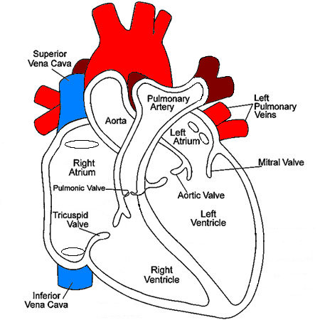
Blood flows to the heart by way of the Superior or Inferior Vena Cava. As blood enters the right atrium it continues across the Tricuspid valve into the right ventricle. With ventricular contraction, the blood then flows out through the Pulmonary valve into the pulmonary arteries to be transported to the lungs. The blood captures oxygen and returns to the left side of the heart by way of the pulmonary veins. As blood enters the left atrium, it continues across the Mitral valve into the left ventricle. When the ventricle contracts, the blood then flows out through the Aortic valve into the Aorta and through the circulatory system.
Blood in the Aorta will flow throughout the circulatory system during systole. During diastole, blood will flow back toward the Aortic Valve and into the coronary arteries supplying the heart muscle with oxygen rich blood.
It is important to realize that blood flows from an area of high pressure to an area of low pressure. As the chambers of the heart relax during diastole, the pressure will become lower in that area and will pull blood from the chamber or vessel that now has higher pressure. As the chamber contracts during systole, the pressure will rise enough to a point where it is higher than the pressure in the next chamber or vessel and blood will flow in that direction. This will be discussed in greater detail when we review Hemodynamics